Computed tomography (CT) is an advanced imaging modality and depending on the area being scanned may be performed under standing sedation or general anaesthesia . Computed tomography has numerous advantages over conventional radiography. CT produces cross-sectional images providing detailed anatomical information where multiple soft tissues and bony structures overlap. CT provides a higher degree of contrast resolution for the soft tissues of the head with the added advantage of being able to performed 3D mulitplanar reconstructed images in sagittal, transverse and dorsal planes.
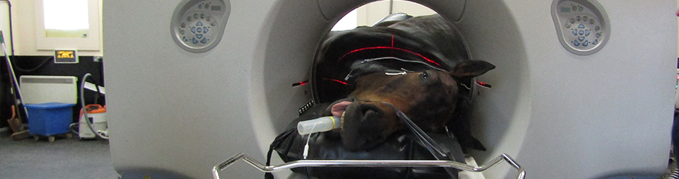
We are pleased to announce at the Liphook Equine Hospital we have installed one of the largest wide bore CT scanner in the UK. The design of our CT suite is unique and purpose built in order to accommodate all shapes and sizes of patients. Our CT scanner has been specifically designed to scan the standing horses head and neck as well as the anaesthetised horse’s limbs. This will enable us to scan more of the horse than ever before. The diameter is 80cm wide so whole body scans will be possible in smaller patients. The platform which the horse stands on is unique to Liphook and able to both lift up and lower down as well as doubling as a bed for the anaesthetised horse.
What is CT?
Computed tomography uses computerised x-rays. A moving gantry scans 360 degrees around the patient which produces thousands of images. These images can then be reconstructed and viewed in many different ways. This imaging modality is very useful when imaging anatomically complex regions such as the head and will help to identify dental, sinus, orbital and temporomandibular joint pathology, diseases of the central nervous and endocrine systems, investigation of head shaking, and traumatic injuries to the head. CT is also a valuable planning tool for surgery.
Whilst our facilities will be available to all inpatients at our hospital we also offer an outpatient referral service for imaging should your veterinarian require our services.