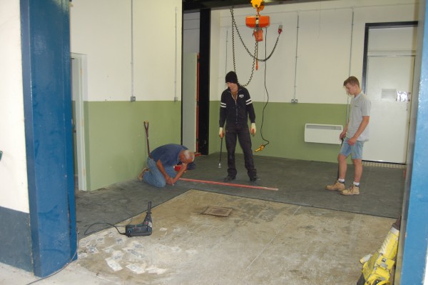
Liphook Equine Hospital is excited to report that we will soon be able to offer a CT scanning service. Building of the new unit has started today and we hope to be able to start scanning horses within 3-4 months.
We are proud to be one of very few equine hospitals in the UK that will have the facilities to carry out CT imaging both in the standing horse and under general anaesthesia.
What is CT?
CT scanning uses a rotating x-ray tube to a take series of x-ray images at high speed circumferentially around the area of interest so giving an image of exquisite anatomical detail allowing the clinician to see subtle changes in bone and soft tissue related to injury and disease. The images can be reconstructed to look at the same piece of anatomy in different orientations and to create a series of detailed slices through the area of interest.
How is CT performed?
CT requires the part of the body to be imaged to be placed into the CT scanner for a short period of time (usually a few minutes). Whether the horse require sedation or general anaesthesia depends upon which part of the horse is being scanned.
Which parts of the horse can be scanned?
The Liphook Equine Hospital plans to install the only large bore (bariatric) CT scanner available for use in horses in the UK. This will allow us not only to scan heads and lower limbs but also mean that we will be able to scan the complete neck of adult horses, the whole body of foals and the proximal parts of the limbs, including stifles.


