Diagnostic Imaging

We operate a modern, multi-modal imaging service for the rapid and effective diagnosis of our patients, including digital radiography, ultrasonography, MRI, CT, scintigraphy, videoendoscopy and overground endoscopy.

About Our Diagnostic Imaging

Alternatively, we offer an advanced imaging service on an outpatient basis for vets who may wish to refer horses for CT or MRI scans and reporting only.
A teleradiology service for referring veterinary surgeons and other referral centres, with detailed and prompt reports is available.

Our Diagnostic services

Computed Tomography (CT)

Computed tomography (CT) is an advanced imaging modality which produces cross-sectional images of a body part, thus providing clinicians with detailed anatomical information.

Our hospital’s wide bore CT scanner is housed in a bespoke, purpose-built CT suite, which allows scans of the head and top part of a horse’s neck to be acquired in the standing, sedated patient or images of the whole neck (to the level of T1/T2) or the whole body in a foal to be acquired under general anaesthesia.


A CT scan generates thousands of images, which can be reconstructed into 3D images and viewed in many different ways, making it easier to visualize complex anatomical structures.

This imaging modality is  particularly useful in horses to image the head to  help to identify dental, sinus, orbital and temporomandibular joint pathology, and in the investigation of causes of  head shaking or assessment of traumatic injuries to the head. 

CT scans of the neck can produce exquisite anatomic detail, allowing recognition of pathologies that cannot be detected by radiography alone. 

By injecting the contrast agent around the spinal cord, the site of spinal cord compression can be identified in ataxic horses (“wobblers”).

Scintigraphy

Nuclear scintigraphy (bone scanning) is a valuable diagnostic technique used in equine lameness and poor performance investigations, complementing other imaging modalities. It involves intravenous injection of Technetium-99m, which variably accumulates in bone and soft tissue depending on their blood supply, the time after injection and bone turnover. A gamma camera detects areas of increased Technetium uptake, identifying sites of injury, inflammation, or altered bone metabolism, often before changes appear on radiographs. The procedure is performed with the horse sedated and standing and is non-invasive, painless, and typically takes 1-3 hours depending on how much of the horse is being scanned. Scintigraphy uses ionising radiation and strict safety regulations apply to the management of these patients, with the horse requiring hospitalisation for 48 hours after injection.

In most cases, a bone scan forms part of a wider orthopaedic evaluation. Scintigraphy is particularly useful in cases where:

Nerve blocks have not localised the site of pain causing lameness or the horse is difficult to safely perform diagnostic analgesia (nerve blocking).

There is multi-limb lameness.
In severe lameness, where a fracture is suspected.
In poor performance cases with no overt lameness.
The area of interest cannot be easily penetrated by radiography (e.g. the back and pelvis).
Lameness has been localised to an area with diagnostic analgesia, but no abnormalities have been detected using other imaging techniques.

Radiography

We offer a full range of mobile and hospital-based radiography services. 

All our equipment uses digital technology, whereby the x-ray beam is converted to an electronic image.  

The digital images can be manipulated, thus enhancing the quality of the images and the systems allow instant results. 

The hospital has two radiography suites with powerful overhead, gantry-mounted units which can be used to obtain high resolution radiographic images of most parts of the horse’s body. 

The hospital-based system generates excellent quality images of the upper limbs, neck and back, which can be difficult to obtain with standard equipment. 

Radiography is also used frequently during surgical procedures, such as fracture repair, and a digital x-ray system in theatre permits instant imaging to help optimise these procedures.

The hospital stores all digital images, using a state-of-the-art computerised database so all images are available almost instantaneously and on-line.

Endoscopy

Endoscopy is used to visualise and evaluate many parts of the horse, including the upper respiratory tract (pharynx, larynx, guttural pouches and trachea), parts of the gastrointestinal tract (oesophagus, stomach, duodenum, rectum and small colon), urinary tract (bladder and urethra) and reproductive tract of mares (vagina, cervix and uterus).

It is an essential part of the evaluation of many horses with poor performance, upper airway obstructions, abnormal respiratory noises, respiratory disease, gastric (stomach) ulcers and many urinary tract problems. 

We have a range of different sized endoscopes at our hospital, which we can use to visualise the different regions of the horse in great detail and take small biopsy samples for laboratory diagnosis.

The endoscopes are linked to a screen (videoendoscopy) which allows the vets and the clients to see the images at the same time and for the images to be recorded.

In some horses with respiratory noise at exercise or demonstrating poor performance, endoscopic examination at rest may appear normal, and yet as the horse is exercised and starts to breathe more deeply, laryngeal or pharyngeal problems become evident. 

Our vets have access to the latest remote dynamic endoscopic system. Significantly, the VetMed remote dynamic endoscope system does not require the use of a high speed treadmill facility and allows endoscopy of a horse when it is being ridden and exercised in a routine manner. 

The equipment is light, relatively unobtrusive and allows the use of the horse’s own tack, which makes it quick and easy to fit to the patient and permits rapid and accurate diagnosis of a multitude of conditions which cause dynamic airway obstruction.

MRI

MRI (magnetic resonance imaging) is an imaging technique that produces highly detailed anatomical images of the bone and soft tissue structures of the area being examined.
Images are produced by placing the body part in a strong magnetic field and applying pulses of radio waves to the part via a radiofrequency coil.
 
A signal is received back via the coil, which is then generated into a detailed image by computer software. 

MRI is a very safe imaging technique as, unlike other imaging techniques (eg. radiography, scintigraphy and CT), it does not involve ionising radiation.

Our hospital was the third equine hospital worldwide to install this pioneering system, and since 2004 we have developed a wealth of experience in using this technique to assist in the diagnosis of equine lameness. 

Our MRI unit was moved to a new, purpose-built suite in 2009 which means that, if required (eg. due to a horse’s temperament), as well as performing MR scans under standing sedation we can also perform scans on horses under general anaesthesia.

The low field MR scanner can be used to image any part of the equine lower limb up to and including the carpus and hock. 

MRI requires the horse to stand still within the magnet with a coil around the area of interest and therefore most horses require sedation as scanning can take between one and three hours. 

The horse’s shoes must be removed before entering the magnet to prevent interference with the strong magnetic field.

MRI is particularly useful for evaluating the bony and soft tissue (eg tendons and ligaments) within the equine foot. 

Our experience has shown that many horses with chronic lameness affecting the front feet have a variety of soft tissue injuries, which cannot be accurately diagnosed using more conventional techniques. 

Furthermore, the increasing use of MRI in horses over the last decade has increased our ability to diagnose and understand many causes of lameness in the fetlock and proximal metacarpal/metatarsal regions.

It is important that the results of MRI are interpreted in conjunction with other clinical findings and diagnostic investigations (eg. regional analgesia and radiography).

Ultrasonography

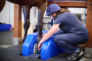
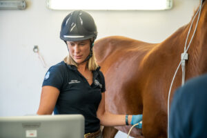
Ultrasonography is a valuable and widely used diagnostic tool in horses. Our equine hospital has several different digital ultrasound machines that can be used for ultrasound scanning of different parts of the body.

It is commonly used for diagnosing and evaluating musculoskeletal conditions, including joint, tendon, muscle and ligament injuries, and can be useful for monitoring the healing of these injuries.

It is also widely used for assessing the abdomen, chest and heart in horses with colic, respiratory or cardiac disease, and monitoring broodmare reproductive cycles and detecting pregnancy. 

We have a range of portable ultrasound machines, as well as diagnostic centre-based machines, the latter being excellent for scanning the more difficult areas such as the chest and abdomen of the horse for the evaluation of heart or lung problems, or as part of examination in colic cases.

Musculoskeletal system
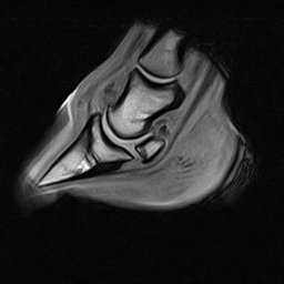
Ultrasonography is used extensively in lameness investigations for the scanning of tendon and ligament injuries, as well as assessing wounds, joint surfaces, fractures and soft tissue swellings. 
Ultrasonography may also be useful for the detection of back and pelvic injuries.

Heart and vascular system
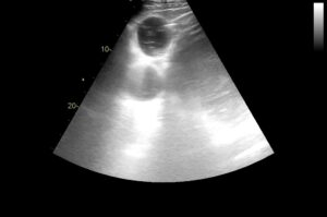
Echocardiogram of the heart is important to assess the chambers and valves of the heart and is invaluable in the assessment of the significance of many types of heart murmur. 

Colour flow Doppler is used to assess dynamic blood flow through different parts of the heart. 

Ultrasonography is also useful for assessing thrombi and peripheral blood vessels.

Abdomen
Ultrasonographic examination of the intestines and other abdominal structures (eg. liver, kidney and spleen) is an important diagnostic tool in the investigation of horses with colic, weight loss or diarrhoea. 

Ultrasound guidance is frequently used to allow safe and precise biopsy of internal structures such as the liver, lungs and kidneys.

Reproductive tract
Ultrasonographic assessment of the ovaries and uterus is important in the management of broodmares to assess the reproductive tract, stage of oestrous cycle and pregnancy diagnosis. 

Regular ultrasound scans of the ovaries are performed (every few hours) of mares undergoing artificial insemination (AI), to ensure that insemination is performed at the optimal time. 

Early pregnancy diagnosis is important to ensure that a mare is not carrying twins.

Thorax
Ultrasonographic assessment of the thoracic cavity, including the lungs, is important in the assessment of horses with pneumonia, lung masses or pleurisy.

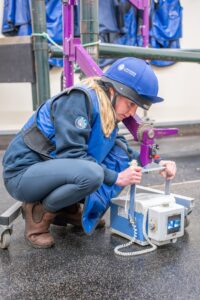
Our veterinary surgeons have access to high quality, mobile digital ultrasonography equipment, meaning that some scans can be done at the horse’s own premises.