Internal Medicine
Our equine internal medicine service is led by RCVS, ECEIM and ACVIM-recognised internal medicine specialists.


Equine Internal Medicine
They provide a highly specialised, 24-hour internal medicine service for the investigation and treatment of a diverse range of internal medical complaints (eg. neurological diseases, respiratory diseases, liver and kidney diseases, diarrhoea, recurrent colic, weight loss, sick neonates).
Horses may be referred from our first-opinion practice or other practices as outpatients for investigation only, or admitted to the hospital for more involved investigations, round-the-clock observation, treatment or critical care.
Animals with contagious or suspected contagious diseases can be admitted into our isolation facilities, and animals requiring critical nursing care admitted into the intensive care unit.
Our large onsite 24-hour laboratory means that rapid analysis and interpretation of blood and other samples is possible.
We also have ultrasonography, endoscopy and other imaging equipment that plays an important role in the investigation of medicine cases.
Some of the investigations available for common disorders are listed below.
Common Disorders
Respiratory diseases
We have a wide range of diagnostic imaging equipment, including gantry-mounted radiography capable of radiographs of the chest, ultrasonography and videoendoscopy, which allows imaging, visualisation and sampling of the respiratory tract, including the paranasal sinuses, trachea, lower airway and guttural pouches.
Samples can be rapidly analysed in our onsite laboratory.
Overground telemetric endoscopy is also available for the diagnosis of upper respiratory tract disorders in exercising horses.
In-house referral of horses to the surgeons for video-endoscopic laser surgery on the upper respiratory tract, under video-endoscopic guidance, tieback, tieforward or thoracoscopy, can be easily arranged.
Cardiac diseases
Heart abnormalities are relatively common in the horse, and can range from being insignificant to life-threatening.
Heart disease may result in heart murmurs, dysrhythmias or exercise intolerance.
Suspected heart problems can be evaluated with a combination of auscultation, echocardiography and colour-flow Doppler ultrasound examination.
Electrocardiographs (ECGs) can be obtained at rest over a 24-hour period and during exercise using a portable recorder.
During this examination, we can also do an exercise tolerance test to see how well they respond to ongoing exercise regimes.
Ocular injuries and diseases
Ocular injuries and diseases in the horse are common, and if inappropriately treated can result in permanent damage to the eye, resulting in loss of vision or even loss of the eye.
Many eye conditions require very intensive treatment, often hourly administration of drops to the eye day and night, which means that they are difficult to treat at home but can be successfully treated in our hospital environment, where vets and nurses are onsite at all hours, throughout the day and night.
We can undertake a full assessment of any eye utilising direct and indirect ophthalmoscopy, as well as ultrasound and tonometry.
Procedures performed by the team include keratopathy, intravitreal injections for uveitis, treatment for glaucoma and ulcer treatment etc. If required, laser treatment for glaucoma and iridial cysts can be organised.
Weight loss investigation
There are many causes of weight loss in the horse. Investigation into causes of weight loss includes physical and dental examination, blood sampling, dynamic absorption testing, faecal analysis and ultrasonography of the abdomen.
Laparoscopic examination of the abdomen can be used to allow visualisation of the abdominal cavity, and when appropriate full exploratory laparotomy of the abdomen can be performed.
Investigation of liver disorders
Liver disease may often present insidiously with non-specific signs, but can be very debilitating.
Initial diagnosis of liver problems is usually based on the results of blood sampling and liver function tests.
However, more specific diagnosis of the type of liver pathology requires ultrasonographic examination of the liver and liver biopsy.
Andy Durham, who leads our internal medicine department, has a particular interest in liver disease and has published and lectured extensively on this subject.
Neurological disorders
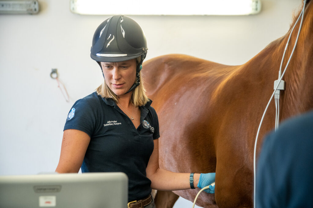
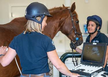
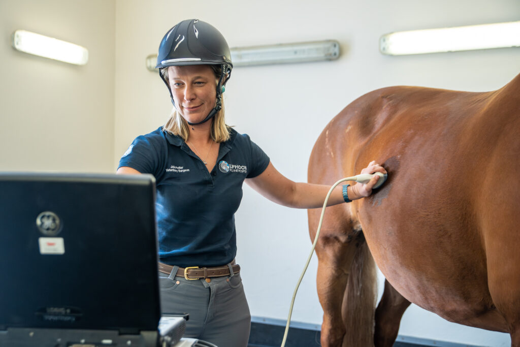
Ultrasonography is a valuable and widely used diagnostic tool in horses. Our equine hospital has several different digital ultrasound machines that can be used for ultrasound scanning of different parts of the body.
It is commonly used for diagnosing and evaluating musculoskeletal conditions, including joint, tendon, muscle and ligament injuries, and can be useful for monitoring the healing of these injuries.
It is also widely used for assessing the abdomen, chest and heart in horses with colic, respiratory or cardiac disease, and monitoring broodmare reproductive cycles and detecting pregnancy.
We have a range of portable ultrasound machines, as well as diagnostic centre-based machines, the latter being excellent for scanning the more difficult areas such as the chest and abdomen of the horse for the evaluation of heart or lung problems, or as part of examination in colic cases.
Musculoskeletal system
Ultrasonography is used extensively in lameness investigations for the scanning of tendon and ligament injuries, as well as assessing wounds, joint surfaces, fractures and soft tissue swellings.
Ultrasonography may also be useful for the detection of back and pelvic injuries.
Heart and vascular system
Echocardiogram of the heart is important to assess the chambers and valves of the heart and is invaluable in the assessment of the significance of many types of heart murmur.
Colour flow Doppler is used to assess dynamic blood flow through different parts of the heart.
Ultrasonography is also useful for assessing thrombi and peripheral blood vessels.
Abdomen
Ultrasonographic examination of the intestines and other abdominal structures (eg. liver, kidney and spleen) is an important diagnostic tool in the investigation of horses with colic, weight loss or diarrhoea.
Ultrasound guidance is frequently used to allow safe and precise biopsy of internal structures such as the liver, lungs and kidneys.
Reproductive tract
Ultrasonographic assessment of the ovaries and uterus is important in the management of broodmares to assess the reproductive tract, stage of oestrous cycle and pregnancy diagnosis.
Regular ultrasound scans of the ovaries are performed (every few hours) of mares undergoing artificial insemination (AI), to ensure that insemination is performed at the optimal time.
Early pregnancy diagnosis is important to ensure that a mare is not carrying twins.
Thorax
Ultrasonographic assessment of the thoracic cavity, including the lungs, is important in the assessment of horses with pneumonia, lung masses or pleurisy.
Our veterinary surgeons have access to high quality, mobile digital ultrasonography equipment, meaning that some scans can be done at the horse’s own premises.
Dermatology
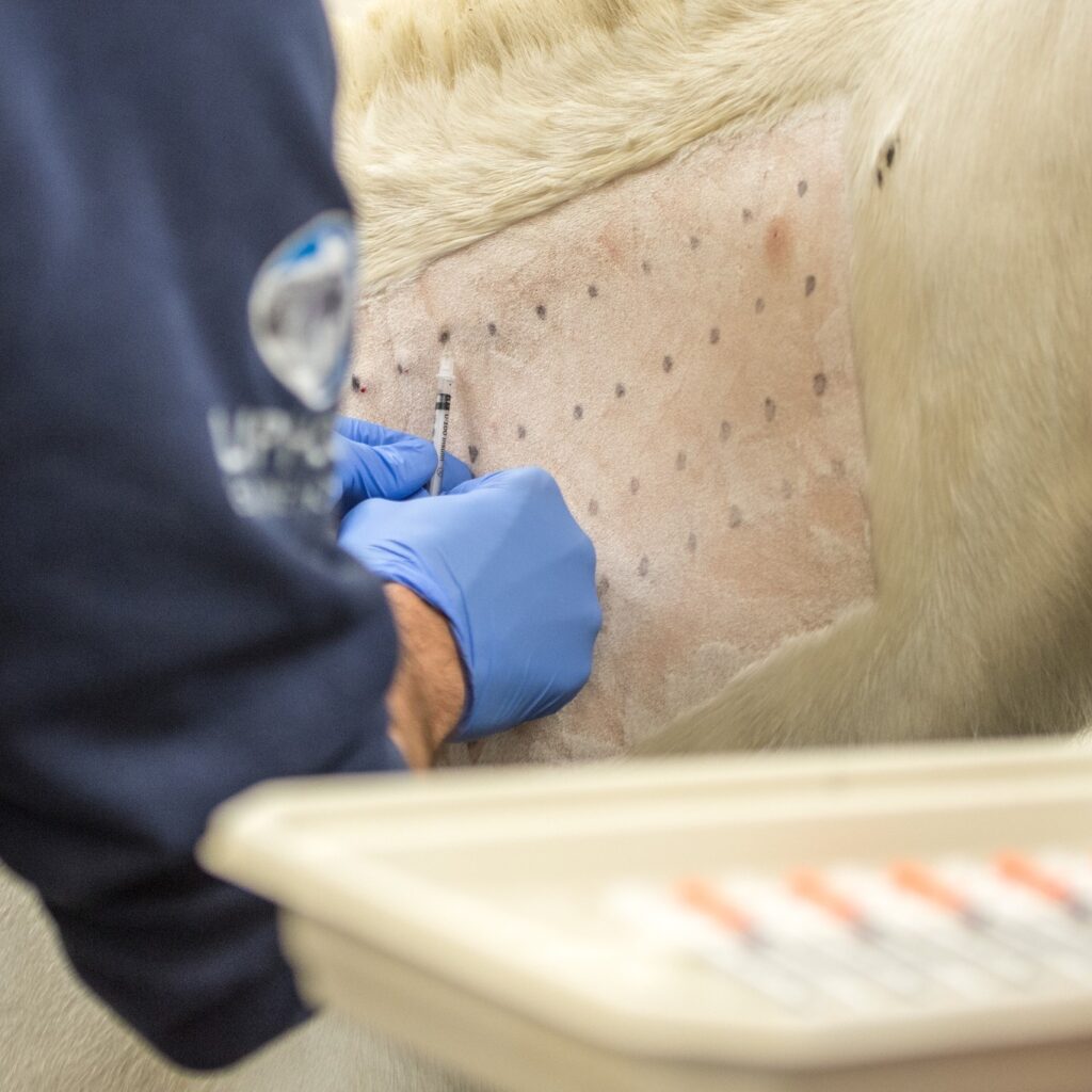
Skin problems in horses are common and can be difficult to diagnose.
Our hospital offers routine appointments and accepts referrals for all types of dermatological conditions, including allergies/immune mediated conditions, bacterial, fungal, parasitic, protozoal or viral diseases, chemical and toxic dermatoses or endocrine disorders.
We are able to perform intradermal skin testing for horses with ongoing pruritus (itchy skin) or skin lesions.
This requires a night of hospitalisation and is the most appropriate way to assess for any allergic disease in the horse.
Often during these examinations, we will also perform skin biopsies, skin scrapes or hair plucks depending on the clinical presentation.
Foal medicine
Sick, premature or dysmature foals require intensive nursing and veterinary care and can deteriorate and die rapidly without such care.
Our intensive care unit is equipped to provide specialist care and 24-hour monitoring of critically ill foals by our experienced clinicians and excellent nursing team.
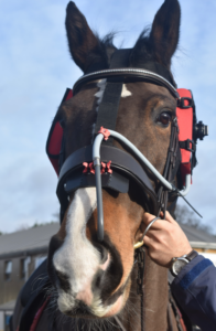
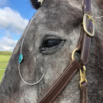
Headshaking Investigation
Headshaking horses show a pattern of signs that can include some or all of the following:
These headshaking signs are sometimes caused by infections, cysts, growths, and inflammation in the head that damage the trigeminal nerve that is responsible for communicating facial sensation to the brain.
More often though, the signs are related to a problem with the function of the nerve itself and we call these cases “trigeminal-mediated headshakers”.
Horses usually develop the condition between the ages of five and 12 years.
It can start rapidly and severely, or it can begin with very mild signs and gradually get worse over months and years.
Some horses only show signs during the spring and summer, and others have signs that persist all year.
The use of computed tomography (CT) has revolutionised our ability to investigate headshaking cases.
It enables us to rule out other diseases, and make a confident and accurate diagnosis of trigeminal-mediated headshaking.
If you suspect your horse is showing headshaking signs, your vets are likely to recommend this important step in the investigation of headshaking signs.
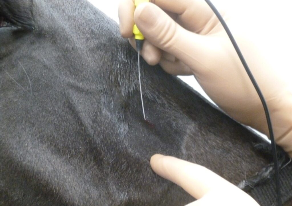
There is an evidence-based, non-invasive treatment option for trigeminal-mediated headshaking cases called percutaneous electrical nerve stimulation therapy.
This PENS treatment has a good success rate in returning horses back to their previous athletic activities and (other than the sedation used during the procedure) has no withdrawal time prior to affiliated competition.
Jamie Prutton has a wealth of experience in managing headshaking cases, and is involved in international research projects in this area.
If you have a suspected headshaker case and would like to find out more then please contact the Hospital for more information: [email protected].